Sonosite M-Turbo Portable Ultrasound Machine
In Stock
| Brand | |
|---|---|
| Condition | |
| Year |
Description
Sonosite M-Turbo Features
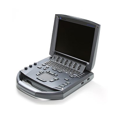
The SonoSite M-Turbo is a portable ultrasound system that is versatile, digital, and software controlled.
This portable and innovative ultrasound system has numerous configurations and feature sets used to obtain and display real-time, high-resolution ultrasonic images. The SonoSite M-Turbo can be used for abdominal, nerve, superficial, vascular, cardiac, venous access, pelvic views, and even in veterinary offices. However used, the M-Turbo ultrasound can optimize the image resolution for high-quality exams. With its clear and sharp contrast, this SonoSite’s ultrasound is known for its great image quality. These features improve the visual detail to help differentiate structures, vessels, and pathology.
The M-Turbo ultrasound has been optimized for real-world environmental challenges. Using its increased processing power the user can get high-quality images for up to 60-seconds. This Ultrasound has a quick boot-up time, backlit keyboard, and is lightweight. It can be carried on site which is Ideal for a variety of ultrasound applications. Even though the M Turbo is a portable ultrasound, it has a 2-hour battery life, depending on the imaging mode and display brightness. The system makes data management easy with a streamlined user-friendly design. It has 2 high-speed USB ports and is Mac and PC friendly.
- 13 compatible transducers
- Lithium-ion battery
- 8 GB internal flash memory
- Integrated speakers
- 3.04 kg
- 30.2 cm diagonal LCD screen
- Premium image quality
- Splash resistant user-interface
- Wireless connectivity
Specifications
Ultrasound Dimensions
- Height: 7.9 cm
- Width: 27.4 cm
- Depth: 30.2 cm
- Weight: 3.04 kg
Display
- Type: Liquid Crystal Display (LCD)
- Size: 26.4 cm
Power
- The system operates via battery or AC power
- Rechargeable lithium-ion battery
- AC: universal power adapter, 100-240 VAC, 50/60 Hz input, 15 VDC output
Onboard Photo and Clip Storage/Review
- 8 GB internal Flash memory storage capability Potential to store 30,000 images or 960 2-second clips
- Clip Store capability (maximum single clip length: 60 seconds)
- Clip Store capability via either the number of heart cycles (using the ECG) or time base Maximum storage in ECG beats mode is 10 heart cycles. Maximum storage in time base mode is 60 seconds
- Cine review up to 255 frame-by-frame images
Measurement Tools, Pictograms, and Annotations
- 2D: Distance calipers, ellipse, and manual trace
- Doppler: Velocity measurements, pressure half-time, auto and manual trace
- M-Mode: Distance and time measurements, heart rate calculation
- User-selectable text and pictograms
- User-defined, application-specific annotations Biopsy guidelines
External Data Management and Wireless
- DICOM Image Management (TCP/IP):
- Print and Store, Modality Work List
- Storage Commit:
- Modality, Perform, Procedure Step
- PC Workstation Image Management (TCP/IP, USB):
- SiteLink Image Manager – allows the transfer, archiving, viewing and printing of high-resolution bitmap images/clips, and batch compression to JPEG on PCs
- Direct writing capability to USB 2.0 mass storage removable media (PC and MAC compatible)
- Supported export formats are: MPEG-4 (H.264), JPEG, BMP, and HTML
- SonoSite Education Key training video compatible
External Video and Audio
- S-video (in/out) to VCR or DVD for record and playback
- RGB or DVI output to an external LCD display
- Composite video output (NTSC/PAL) to VCR or DVD, video printer or external LCD display
- Audio output
- Integrated speakers
H-Universal Stand and Peripherals
- Transducer and gel holders
- Optional Triple Transducer Connect (TTC) to quickly activate transducers electronically
- Optional foot switch
Optional Peripherals
- Printers: Medical-grade black and white or color
- External storage devices: Medical-grade DVD
- External data input devices: Barcode reader ECG module: 3-lead ECG – works with standard ECG leads and electrodes External analog ECG input also available
- USB barcode reader
Broadband, Multifrequency Imaging
- 2D / Tissue Harmonic Imaging / M-Mode
- Velocity Colour Doppler / Colour Power Doppler
- PW, PW Tissue Doppler and CW
- Doppler angle, correct after freeze
Image Processing
- SonoADAPT Tissue Optimization
- SonoHD Imaging Technology
- Advanced Needle Visualisation (SonoMBe Imaging)
- Dual Imaging, Duplex Imaging, 2x pan/zoom capability, Dynamic range and gain
Transducer Types
- Broadband and Multifrequency:
- Linear Array, Curved Array, Phased Array, Multiplane TEE and Micro-Convex
- Single Frequency:
- Cardiac Static Pencil
Application Specific Calculations
- OB/GYN/Fertility: Diameter/ellipse measurements, volume, ten follicle measurements, estimated fetal weight, established due date, gestational age, last menstrual period, growth charts, user-defined tables, multiple user-selectable authors, ratios, amniotic fluid index, patient report, humerus and tibia measurement and charts
- Vascular: Diameter/ellipse/trace measurements, volume, volume flow, percent diameter and area reduction, Lt/Rt CCA, ICA, ECA, ICA/CCA ratio, time average mean (TAM), peak trace, ICA/CCA ratio, angle correction, patient report
- CIMT (Carotid Intima Media Thickness): Embedded SonoCalc IMT software (optional) – automatic edge detection with mean and maximum thickness reporting
- Cardiac: Automated Cardiac Output package and patient report including ventricular, aortic and atrial measurements; ejection fraction, volume measurements, Simpson’s rule, continuity equation, pressure half-time and cardiac output; PA AT, TV E, A, PHT, TVI, MV time, Pulm Veins
- Transcranial Doppler (TCD): Complete TCD package including Time Average Peak (TAP)
Compatible Transducers
- L38xi
- HFL38x
- HFL50x
- L25x
- C11x
- C60x
- ICTx
- P21x
- P10x
- SLAx
- TOEx/TEEx
- D2x
- L52x
Additional information
| Brand | |
|---|---|
| Condition | |
| Year |


